
3D head-shape screening for primary care

SoftSpot3D

An iPad becomes a 3D head scanner for infants.

One 360° scan gives a full 3D model of a baby’s head and an automatic head-shape report, in a regular exam room. We’re building SoftSpot3D to make head-shape screening in primary care more objective and easier to access.

Validated against a medical-grade 3D system in 143 infants at Children’s National Hospital and Wake Forest School of Medicine. Supported by an NIH SBIR award. Patent pending. See the research

SoftSpot3D is not yet commercially available.

Why SoftSpot3D

Bring 3D head-shape measurement to primary care

Flat head and craniosynostosis are easiest to treat in the first months of life. Early screening usually happens at routine well-child visits, and it is mostly visual. 3D scanners measure head shape objectively, but they fill a room and cost too much for a pediatric office, so they sit in specialty centers.

SoftSpot3D isn’t built to make helmets. It’s built to make head-shape screening more objective and more accessible, for earlier detection and better-informed referrals.

= camera

Room-sized 3D scanner

A frame of synchronized cameras (orange) in a dedicated room. Accurate, but bulky, expensive and fixed in place.

  • Dedicated room
  • Fixed in place
  • Specialty centers
depth sensor

SoftSpot3D

An iPad with a clip-on depth sensor. Portable and low-cost, made for a routine visit.

  • Any exam room
  • Portable
  • Primary care
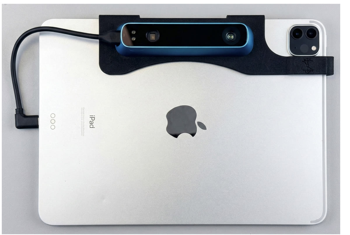
An iPad Pro with a Structure Sensor depth camera mounted on the back in a black bracket, connected by a short cable.
iPad Pro with a Structure Sensor mounted on the back.
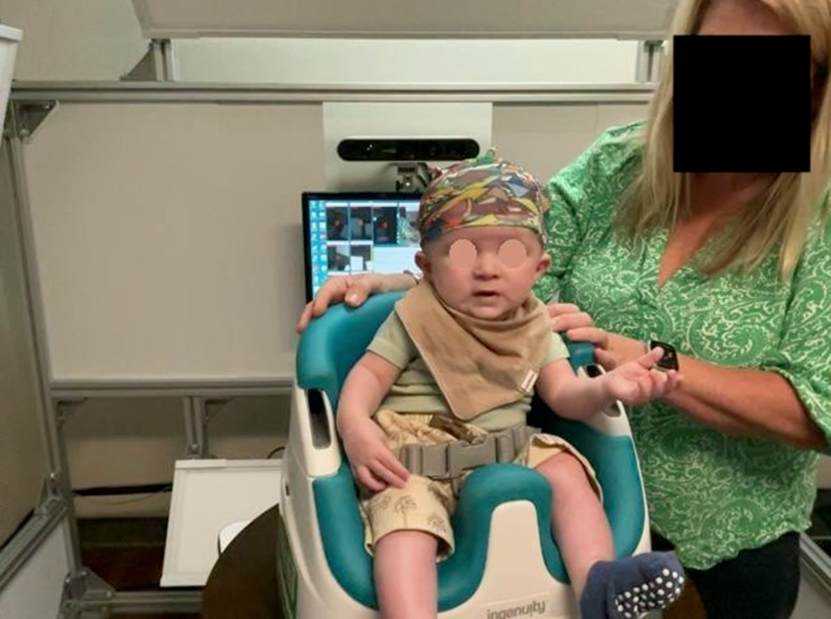
A SoftSpot3D scan session: an infant in the patterned cap sits in the baby seat on the turntable, facing the depth sensor, while an operator steadies the seat. Faces are masked.
A scan session in the study: the patterned cap, the baby seat on the turntable and the depth sensor. Faces are masked.

The hardware

An iPad, a depth sensor and a cap

iPad Pro
Runs the SoftSpot3D app, records the scan and guides the operator with live distance and coverage feedback.
Structure Sensor
A structured-light depth camera clipped to the iPad. Its depth error is about 0.5 mm at 40 cm, where the iPad’s built-in LiDAR can be off by more than 10 mm.
Patterned cap
A colored nylon cap gives the camera features to track. Its exact placement doesn’t matter.
Turntable seat
The baby sits on a turntable and turns once in front of the iPad.

How it works

From one scan to a 3D head model and report

  1. 01

    Put on the cap

    A patterned nylon cap gives the camera features to follow. It is only there for tracking, so its exact position doesn’t matter.

  2. 02

    Scan once around

    The baby sits on a turntable for one 360° turn in front of an iPad with a depth sensor. The app shows distance and coverage as you go.

  3. 03

    Rebuild the head in 3D

    The software follows the baby’s natural head movement, corrects for it, and builds a closed 3D model of the head.

  4. 04

    Read the report

    Head circumference, cranial index, cranial vault asymmetry and its index, and cranial volume, with top, left, right and back views.

What it captures

A 3D head model, landmarks and measurements

3D head model

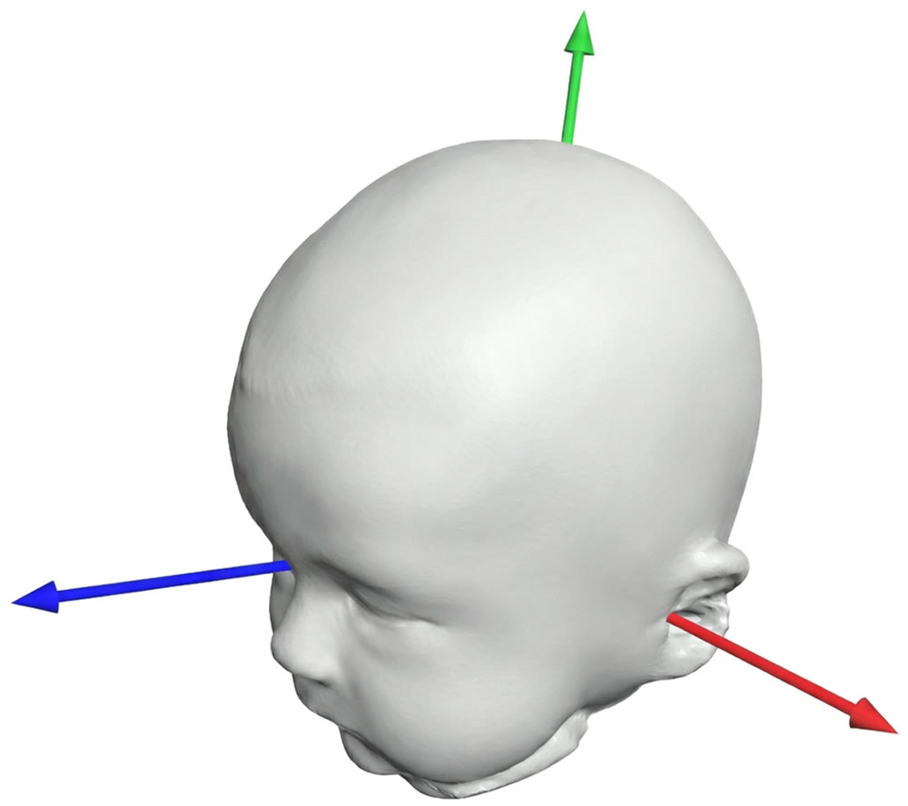
A closed 3D model of the whole head, rebuilt from one turn even when the baby moves. This is a real SoftSpot3D reconstruction: drag it to look around.

3D model of an infant's head with three axes defined from the nasion and both tragions.

Landmarks

The nasion and both tragions set a standard coordinate system, so every head is measured the same way. Landmarks can be placed by hand or found automatically.

Measurements and report

  • HCHead circumference
  • CICranial index
  • CVACranial vault asymmetry
  • CVAICranial vault asymmetry index
  • VolCranial volume
  • MoreCraniosynostosis risk, with the likely affected sutureIn research

Measured on the plane where head circumference is largest above the ears, then compiled into an automatic report with top, left, right and back views. In research: a machine-learning model trained on 3D head shapes to flag shapes that look like craniosynostosis rather than positional flattening, to support earlier, better-informed referrals. See the research

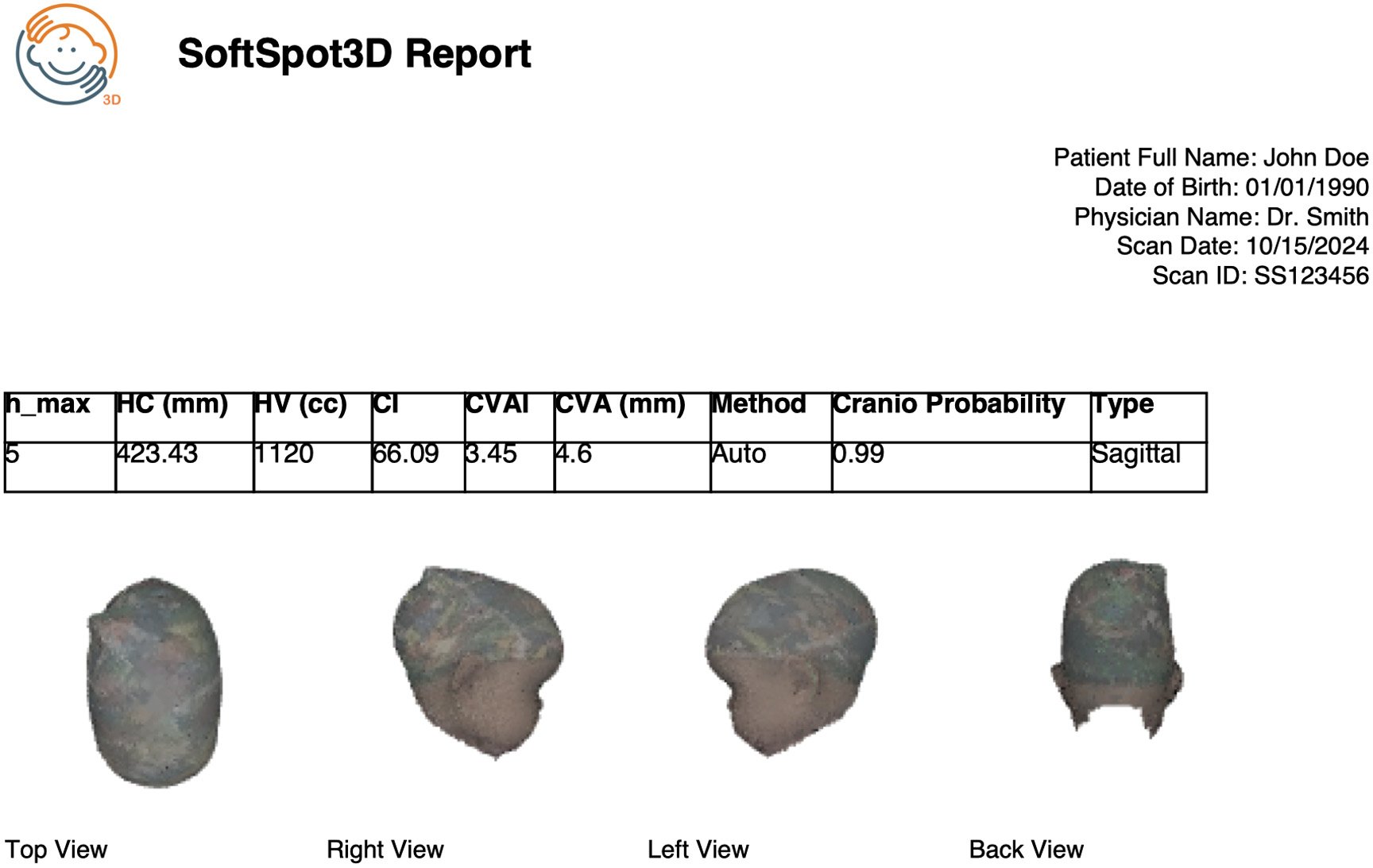
Example SoftSpot3D report: a header with placeholder patient details, a table of head circumference, head volume, cranial index, CVAI, CVA, craniosynostosis probability and type, and top, right, left and back views of the 3D head model.

Example report

Generated automatically from one scan, with no manual steps: head circumference, head volume, cranial index, CVAI and CVA, a craniosynostosis probability with the suspected type (in research), and top, right, left and back views of the head. The patient details shown are placeholders.

Hardware, setup, 3D model, landmark and report images: Wu et al., Journal of Medical Imaging 13(5), 054502 (2026).

Research

Published research

This work was supported by an NIH SBIR award from the National Institute of Dental & Craniofacial Research (Award No. SB1DE034606).

Patent pending: “System and Methods for Motion Robust 3D Reconstruction and Measurement of Body Parts” (U.S. Patent Application No. 63/631,776).

Journal of Medical Imaging · 2026

SoftSpot3D: prospective study of mobile 3D reconstruction for infants’ cranial conditions

Wu, Kocabalkanli, Aalamifar, Linguraru, et al.

Compared SoftSpot3D with 3dMD in 143 infants at Children’s National Hospital and Wake Forest School of Medicine. The cranial surface was within 2.94 mm on average, and head-shape indices agreed closely.

Read the paper

The Cleft Palate Craniofacial Journal · 2026

Machine Learning-Based Identification of Craniosynostosis From Nonsynostotic Cranial Deformities

Kocabalkanli, Wu, Blondin, Mantilla-Rivas, et al.

Uses machine learning on 3D head shapes to tell single-suture craniosynostosis from nonsynostotic head shapes and to identify the affected suture, to help pediatric providers tell the two apart early.

Read the paper
Summary of the SoftSpot3D study in the Journal of Medical Imaging
infants measured at two hospitals
143
average distance between the SoftSpot3D and 3dMD cranial surfaces
2.94 mm
mean absolute error in cranial index (CI)
2.18
mean absolute error in cranial vault asymmetry index (CVAI)
2.16

Why it matters

Positional flat head affects an estimated 16–20% of U.S. infants in the first year, and craniosynostosis needs urgent surgical evaluation. Both respond best to care within a narrow 4-to-6-month window, yet primary-care screening is mostly visual. 3D scanners can measure head shape objectively, but they are costly and fixed in place.

What the study did

A prospective study at Children’s National Hospital and Wake Forest School of Medicine scanned each infant with SoftSpot3D on an iPad and with 3dMD, a medical-grade stereophotogrammetry system used as the reference. Of 194 infants scanned, 157 (80.9%) were reconstructed and 143 had complete measurements.

What it found

The SoftSpot3D cranial surface was on average 2.94 mm from the 3dMD surface. Head-shape indices agreed closely, with little systematic bias: −0.39 for cranial index and 0.44 for cranial vault asymmetry index. Automatic landmarking performed about as well as manual expert landmarking.

What it means

The errors in cranial index and asymmetry index sit well within the ranges that separate clinical severity levels. The authors conclude that a mobile 3D scan can bring objective head-shape assessment into routine pediatric care, with a structured report generated automatically.

Difference from 3dMD, 143 infants
MeasurementMean ± SD
Cranial surface distance (Hausdorff)2.94 ± 1.32 mm
Cranial index (CI)2.18 ± 1.63
Cranial vault asymmetry index (CVAI)2.16 ± 1.90
Cranial vault asymmetry (CVA)3.10 ± 2.68 mm
Head circumference (HC)10.97 ± 9.56 mm

Surface distance is the average Hausdorff distance; the other rows are mean absolute errors.

Bo Wu, Can Kocabalkanli, Fereshteh Aalamifar, Marius George Linguraru, Athena Zhang, Robert Francis Keating, Angela Myers, Mario Blondin, Mary Kathryn Hederick, Christopher Michael Runyan, Gary Francis Rogers, Reza Seifabadi. Journal of Medical Imaging 13(5), 054502, published October 16, 2026. doi:10.1117/1.JMI.13.5.054502

Supported by the National Institute of Dental & Craniofacial Research of the NIH under Award No. SB1DE034606. Five of the authors are PediaMetrix employees; PediaMetrix owns SoftSpot3D.

Collaborate with us

SoftSpot3D is still in development. We’d like to hear from pediatric and primary care practices, craniofacial teams and research groups who want to help bring 3D head-shape measurement into routine care.

Measuring head shape today? SoftSpot® is FDA-cleared and on the market. Explore SoftSpot Pro

SoftSpot3D is not yet commercially available. Study results are from research and are not a diagnosis.