
For healthcare providers only
CranioSpot™
See head-shape patterns associated with craniosynostosis — from the photos your SoftSpot session already takes.


Sign in with your SoftSpot provider account — in the app or on the web portal. CranioSpot results appear on the provider report only, never on the parent report.
Not a medical device. CranioSpot gives a recommendation to support clinical judgment; it does not diagnose or rule out craniosynostosis.
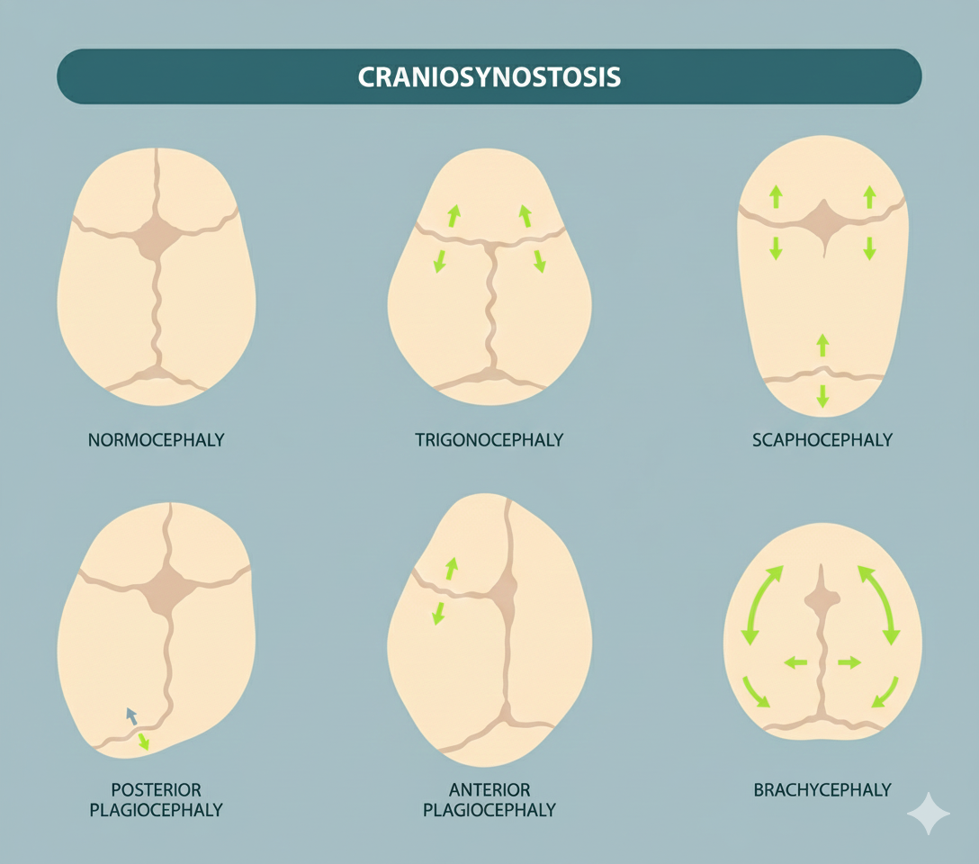
Types of craniosynostosis
Craniosynostosis head shapes, seen from above
Each shape below comes from an early-fused suture — craniosynostosis, not positional flattening — shown beside a typical head for comparison. CranioSpot reviews the head outline for patterns like these.
For the parent-facing explanation of how the two differ, see craniosynostosis vs. positional flat head.
Typical head
ReferenceTypical proportions, all sutures open.
Sagittal synostosis
Long and narrow. The most common single-suture form.
Metopic synostosis
Narrow, pointed forehead with a midline ridge.
Unilateral coronal synostosis
One side of the forehead flattened.
Bilateral coronal synostosis
Short and wide.
Unilateral lambdoid synostosis
Back flattened on one side. Rarest, and easily mistaken for positional flattening.
Schematic illustrations. Real cases vary and can overlap.
Clinical background · SoftSpot Resources
Craniosynostosis in Babies
Suture types, signs, and how craniosynostosis differs from positional flattening — ready to share with families.
How it works
No extra capture. No new workflow.
Step 1
Capture as usual
Auto Capture guides the framing — the outline turns green when the head is aligned. Same photos as any SoftSpot session.
Step 2
CranioSpot reads the outline
Once the photos upload, AI reviews each one's head outline for shape patterns.
Step 3
Review the provider report
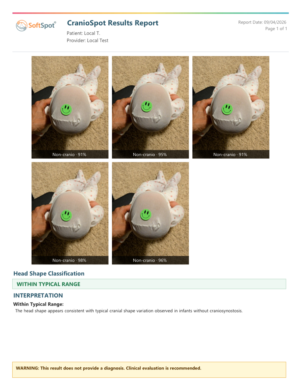
A result label and recommended next step, with the photos — in the app or on the web portal.
Head shape consistent with typical infant variation.
No action prompted by this result.
Not a clearance — pursue any clinical concern regardless of the label.
Head shape shows features that can be associated with craniosynostosis.
Flagged for clinical review alongside examination.
Not a diagnosis — positional flattening can also produce this label.
Sample output
The CranioSpot results report
Questions
Common questions
Research
Publications behind CranioSpot
- SPIE Medical Imaging 2021 · Computer-Aided DiagnosisPhotographic cranial shape analysis using deep learningYektaie, M.A., Ghasemi, Z., Hezaveh, S.H., Aalamifar, F., Seifabadi, R., Linguraru, M.G.Read the paper
- Pediatric Academic Societies Meeting 2023Differentiating Synostotic vs Non-Synostotic Cranial Conditions from Photographs using Deep Learning: a Retrospective StudyKocabalkanli, C., Rahmani, S., Aalamifar, F., Linguraru, M.G., Seifabadi, R.View the abstract