
Parent measurement guide
How Is a Baby's Head Shape Measured? CI and CVAI Explained
Looking at a baby's head can help parents notice possible flattening. Objective measurements go a step further by putting numbers to overall head proportions and differences between the two sides, making head shape easier to describe and track.
Reviewed by the SoftSpot Clinical & Scientific Team
Why measure a baby's head shape?
Parents often begin with a visual observation: one side of the back may look flatter, the whole back may look broad, or the ears may not appear aligned from above. These observations are useful because they identify something to discuss. They cannot reliably show how large a difference is, whether it is changing, or what caused it.
Routine infant care includes a physical assessment of head and skull shape. Visual examination remains important, but it does not provide a numerical measure of head proportion or asymmetry. Objective measurements such as CI and CVAI can complement routine clinical assessment and make it possible to track those dimensions consistently over time.
A baby head shape measurement uses defined dimensions and a repeatable method. Instead of relying on memory or photographs taken at different angles, measurements create a numerical description that can be compared over time. That distinction matters when parents and clinicians want to know whether proportions are staying similar as the head grows or changing between checks.
Objective does not mean diagnostic. The same number can have different meaning depending on age, measurement method, head growth, neck movement, birth history, and the rest of the examination. If you are first trying to understand what you see, read Is My Baby's Head Shape Normal? before focusing on the numbers.
What measurements are used?
Most numerical descriptions begin with a top view of the head and a set of consistent landmarks. The exact technique can differ among tools and clinical systems, which is one reason results should be compared using the same method whenever possible.
Head width and length
Width describes the maximum side-to-side dimension. Length describes the front-to-back dimension. Together, they describe overall proportion.
Diagonal measurements
Two lines cross the head at matching angles on opposite sides. Comparing their lengths provides information about cranial asymmetry.
Cranial/Cephalic Index
Cranial Index, also called Cephalic Index, relates head width to head length and is commonly abbreviated CI.
CVA and CVAI
Cranial Vault Asymmetry is the difference between diagonal lengths. Cranial Vault Asymmetry Index expresses that difference relative to head size.
Head circumference is also routinely followed as part of infant growth, but circumference alone does not describe width-to-length proportion or side-to-side asymmetry. A provider may consider several measurements together rather than treating one value as a complete picture.
What is Cranial Index (CI)?
Cranial Index describes how wide the head is relative to how long it is. You may also see the term Cephalic Index; in infant head-shape discussions, both are commonly abbreviated CI. A CI baby head result is a proportion, not a direct measurement in inches or millimeters.
Cranial Index formula
CI = head width ÷ head length × 100
For a simple example, a relatively wider head produces a higher ratio than a relatively narrower head of the same length. The calculation makes proportions easier to compare across different head sizes. It does not reveal why the proportion looks that way, and CI alone should never be presented as a diagnosis or treatment recommendation.
What is CVAI?
CVAI stands for Cranial Vault Asymmetry Index. It begins with two diagonal dimensions measured across the head at corresponding angles. When one diagonal is longer than the other, their difference describes the amount of asymmetry. CVAI relates that difference to the size of the head and reports it as a percentage.
A related value, Cranial Vault Asymmetry (CVA), is the difference between the two diagonal measurements, usually expressed as a distance. CVAI is an index rather than a raw distance. That can be helpful when following an infant whose whole head is growing.
There is not one universal CVAI classification used identically by every device, clinic, study, and health system. Diagonal angle, landmarks, calculation details, and named categories can vary. This article therefore does not provide a severity cutoff. The result should be interpreted according to the method used and in the context of a qualified clinical evaluation.
What Do CI and CVAI Numbers Mean?
CI (Cranial Index) shows how wide versus long the head is. CVAI (Cranial Vault Asymmetry Index) shows how symmetrical the head is from left to right.
What CI describes
width ÷ length
A higher CI describes a relatively wider, shorter head shape (brachycephaly); a lower CI describes a longer, narrower one. Published normal ranges for CI differ between clinics, devices and studies, so a single cutoff is not quoted here — ask which reference range your provider uses.
CHOA CVAI reference
<3.5%
The CHOA guideline presents a normal CVAI threshold below 3.5%. Higher CVAI reflects greater left-to-right cranial asymmetry.
The CVAI threshold above is from the Children's Healthcare of Atlanta (CHOA) plagiocephaly severity scale, which grades asymmetry by CVAI and does not set a CI range.
CI and CVAI are measurements, not diagnoses. Reference ranges and severity classifications should be considered in clinical context and do not by themselves determine whether treatment or helmet therapy is needed.
Wondering what to try while you track the numbers? Read how to help a baby's flat head without a helmet.
Plagiocephaly vs. brachycephaly measurements
Positional plagiocephaly generally refers to a pattern with greater flattening on one side. Because the pattern is asymmetric, diagonal differences, CVA, and CVAI are particularly relevant when a clinician describes or follows it. A plagiocephaly measurement still does not replace examination of the entire head, sutures, face, ears, and neck movement.
Brachycephalic head shape generally refers to a proportion that is relatively wide from side to side and short from front to back, often with broader flattening across the back. CI helps describe that overall width-to-length proportion. Plagiocephaly and brachycephaly can occur together, so CI and CVAI may both appear in one report.
Parents searching how to measure a flat head baby should be cautious with online charts and home-made cutoffs. A number cannot establish cause, rule out craniosynostosis, or determine care. Neck tightness or a preferred turning direction may also be relevant; the Resource Center includes an existing torticollis resource.
How are these measurements traditionally obtained?

Manual calipers
Trained clinicians can use anthropometric calipers to measure width, length, and diagonals at defined landmarks. Calipers are practical and widely used, but technique matters. Infant movement, hair, landmark placement, instrument angle, and differences between measurers can affect repeatability.
3D imaging and surface scanning
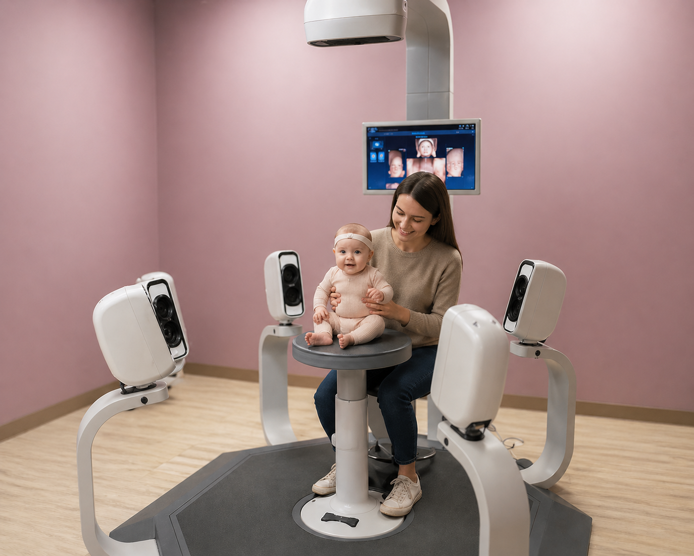
Three-dimensional photography or surface scanning captures the outside contour of the head and allows dimensions to be calculated digitally. These systems can provide detailed and repeatable surface information but may require specialized equipment, space, training, and an in-person visit. They are different from medical imaging used to examine structures inside the skull.
Standardized photography and other validated methods may also be used. Whichever approach is chosen, comparing results is most meaningful when capture position, landmarks, formulas, and measurement method remain consistent.
Measuring with SoftSpot®

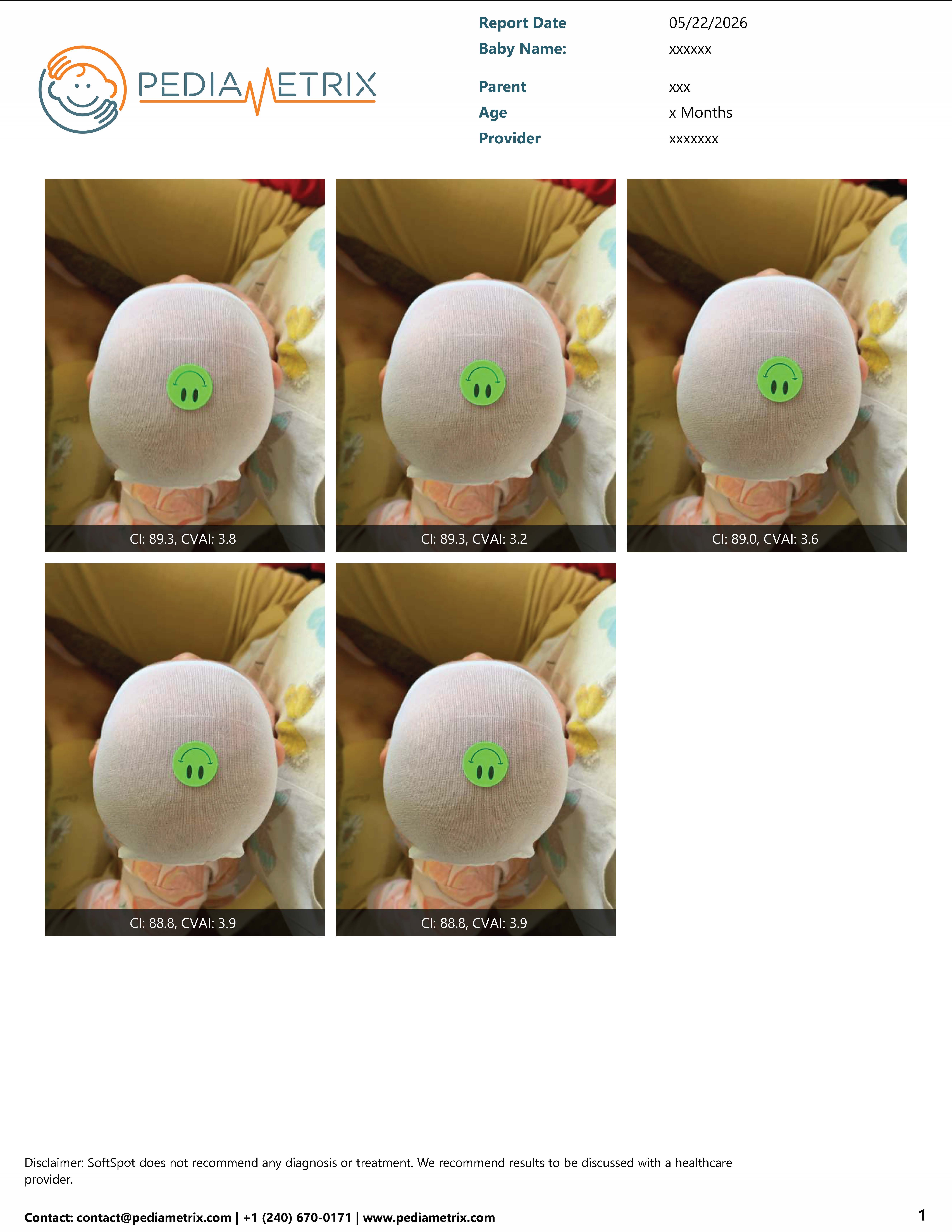
SoftSpot® is an FDA-cleared cranial measurement tool that uses a smartphone-based workflow to obtain objective infant head-shape measurements, including CI and CVAI. Guided top-view capture makes it possible to document measurements without specialized 3D scanning equipment in the home.
The measurement report provides a consistent numerical record that parents can use to track head shape over time and bring to conversations with a healthcare provider. Convenience does not change the role of the tool: SoftSpot measures physical head-shape characteristics. It does not diagnose plagiocephaly, determine treatment, assign a universal clinical severity, or decide whether a helmet is needed.
For practical setup and capture guidance, use the existing Getting Started with SoftSpot® guide.
Understanding your SoftSpot report
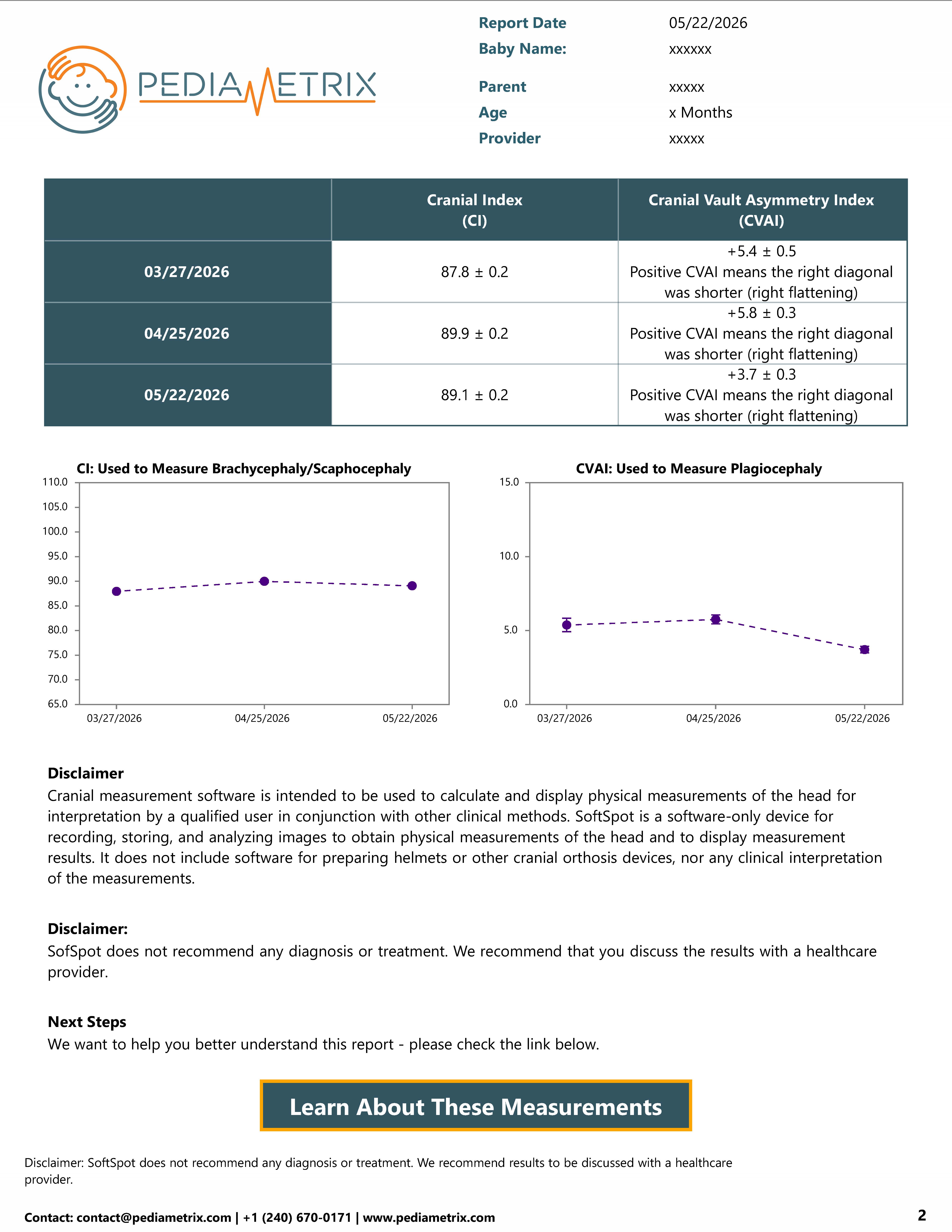
Your report organizes the captured images and head-shape measurements for review. Rather than repeating those details here, visit How to Read Your SoftSpot® Report for the existing step-by-step explanation of its measurement layout.
When should I talk to my baby's healthcare provider?
Ask at any well-child visit if you notice a flat area, a change in head shape, uneven ears or forehead, a persistent head tilt, or a strong preference for looking one way. Contact the provider sooner if neck movement seems limited, the shape changes quickly, you feel an unusual ridge, or you have any concern about growth or development.
Bring observations, consistent photos, or measurement reports if available. A pediatrician or qualified healthcare provider can examine the skull and sutures, review head growth and birth history, assess neck movement, and decide whether follow-up or referral is appropriate. Measurements support that conversation; they do not replace it.
Continue established safe-sleep guidance: place your baby on their back for every sleep on a firm, flat surface. Do not use head-shaping pillows or change sleep position because of a measurement. Tummy time and varied positioning are for awake, directly supervised periods unless your child's clinician gives individualized instructions.
Frequently asked questions
How do you measure a baby's head shape?
Infant head shape can be described using width, front-to-back length, and diagonal measurements taken with a consistent method. Clinicians may use calipers, standardized photographs, or 3D surface imaging. Measurements are interpreted together with physical examination, age, growth, and medical history.
What is Cranial Index (CI)?
Cranial Index, also called Cephalic Index, describes head width relative to head length. It is calculated as head width divided by head length, multiplied by 100. CI describes proportion but does not diagnose a condition by itself.
What is CVAI?
Cranial Vault Asymmetry Index, or CVAI, uses the difference between two diagonal head measurements to describe cranial asymmetry relative to head size. Methods and classification systems vary, so a result needs appropriate clinical context.
What is the difference between CI and CVAI?
CI compares overall head width with length. CVAI compares diagonal measurements to describe how different the two sides are. They describe different aspects of infant head shape and neither is a stand-alone diagnosis.
Can I measure my baby's head shape at home?
Parents can observe shape at home, but improvised tape or caliper measurements can be inconsistent and should not be used to diagnose or choose treatment. An FDA-cleared measurement tool such as SoftSpot can provide objective measurements through its guided smartphone workflow. Discuss results and concerns with a qualified healthcare provider.
How is plagiocephaly measured?
A clinician evaluates the whole head and may compare diagonal dimensions using Cranial Vault Asymmetry or CVAI. Visual examination, neck movement, growth, age, and the possibility of other causes remain important; a measurement alone does not diagnose plagiocephaly.
Can head-shape measurements tell whether my baby needs a helmet?
No. Measurements are only one part of a clinical assessment. Decisions about observation, physical therapy, a cranial orthosis, or other care depend on age, growth, examination, cause, change over time, and other clinical findings. A qualified healthcare provider should make treatment recommendations with the family.
References
- U.S. Food and Drug Administration. 510(k) Summary K212045: SoftSpot Cranial Measurement Software.
- American Academy of Pediatrics, HealthyChildren.org. When a Baby's Head Is Misshapen: Positional Skull Deformities.
- Johns Hopkins Medicine. Deformational Plagiocephaly.
- Hagan JF Jr, Shaw JS, Duncan PM, eds. Bright Futures: Guidelines for Health Supervision of Infants, Children, and Adolescents. 4th ed. American Academy of Pediatrics. 2017.
- Feijen MMW, Schuckman M, Habets E, van der Hulst RRWJ. Positional plagiocephaly and brachycephaly: is there a correlation between subjective and objective assessment of cranial shape? J Craniofac Surg. 2012;23(4):998-1001. PMID: 22777451.
- Methods to Diagnose, Classify, and Monitor Infantile Deformational Plagiocephaly and Brachycephaly: A Narrative Review. Journal of Chiropractic Medicine. 2015.
- Nieberle F, et al. Direct Anthropometry Overestimates Cranial Asymmetry—3D Digital Photography Proves to Be a Reliable Alternative. Diagnostics. 2023.