
Clinical resource for pediatric providers
A Practical Guide to Infant Head Shape Assessment for Pediatric Providers
A scope-conscious framework for recognizing positional cranial deformity, adding objective measurements to clinical observations, following change over time, and communicating when referral is appropriate.
Reviewed by the SoftSpot Clinical & Scientific Team
1. Why infant head shape belongs on the clinical radar
Pediatric PTs and OTs may see infants because of torticollis, positional preference, motor delay, postural asymmetry, or developmental concerns. Lactation consultants often observe head and neck positioning closely during feeding. Chiropractors and other infant-care professionals may meet families who already have questions about a flat spot or turning preference.
These touchpoints can make pediatric professionals useful observers between well-child visits. That does not give every profession the same authority to diagnose or treat plagiocephaly. The appropriate role may be examination and management, structured observation, objective documentation, parent education, communication with the pediatrician, or referral—depending on discipline and jurisdiction.
2. What should providers look for?
Typical symmetry
Plagiocephaly
Brachycephaly
Observe the head from above and consider the posterior contour, forehead, ears, facial alignment, and neck movement together. Atypical shape, palpable ridging, unusual facial or orbital findings, progressive deformity, or concern for craniosynostosis warrants appropriate medical evaluation rather than positional assumptions.
3. Visual assessment vs. objective measurement
Observation remains essential. It provides information about contour, associated features, posture, movement, and the broader clinical presentation. What it does not provide is a numerical baseline.
Objective measurement can complement visual and clinical assessment by providing reproducible numerical data that can be followed over time.
A consistent measurement can help answer: How asymmetric is the head? Is it proportionally wider and shorter? Is the shape changing? Is asymmetry improving, stable, or worsening? Can the trend be documented across visits? Reproducibility still depends on consistent landmarks, positioning, technique, and tool use.
4. Understanding CI and CVAI
Cranial Index (CI)
Maximum cranial width divided by front-to-back length, multiplied by 100. Also called Cephalic Index, CI describes width-to-length proportion and is relevant when evaluating brachycephalic or dolichocephalic proportions.
Cranial Vault Asymmetry Index (CVAI)
The absolute difference between paired diagonal measurements divided by the larger diagonal, multiplied by 100. CVAI expresses side-to-side cranial asymmetry relative to head size.
The Children's Healthcare of Atlanta (CHOA) scale is one published classification system for CVAI and cranial proportion. Its ranges should be attributed to that guideline—not treated as universal thresholds. Systems, landmarks, and clinical pathways vary, and a category does not independently establish diagnosis or treatment.
For formulas and interpretation context, see Understanding Your SoftSpot Report and How Is a Baby's Head Shape Measured?.
What Do CI and CVAI Numbers Mean?
CI (Cranial Index) describes how wide the head is relative to its length. CVAI (Cranial Vault Asymmetry Index) describes the degree of left-to-right diagonal asymmetry.
What CI describes
width ÷ length
A higher CI describes a relatively wider, shorter head shape; a lower CI describes a longer, narrower shape. Published reference ranges vary among clinics, devices, populations, and studies, so document which reference system and measurement method your practice uses.
What CVAI describes
|diagonal A − diagonal B| ÷ larger diagonal
A higher CVAI describes greater measured left-to-right asymmetry. In the CHOA scale, CVAI below 3.5% is categorized as normal; that is one named classification system, not a universal diagnostic threshold.
The threshold above is specific to the CHOA plagiocephaly severity scale. CHOA does not provide a CI range in that document, and its CVAI categories should not be presented as universal thresholds.
CI and CVAI are measurements, not diagnoses. A value or category does not establish etiology, rule out craniosynostosis, or independently determine therapy, referral, or helmet candidacy.
How are CI and CVAI measured? Calipers, 3D scans and apps

Manual calipers (craniometers)
Trained clinicians can measure width, length, and diagonals at defined anthropometric landmarks with a cranial caliper, also called a craniometer. Calipers are practical and widely used, but repeatability depends on landmark placement, instrument angle, infant movement, hair, and examiner technique.
3D imaging and surface scanning
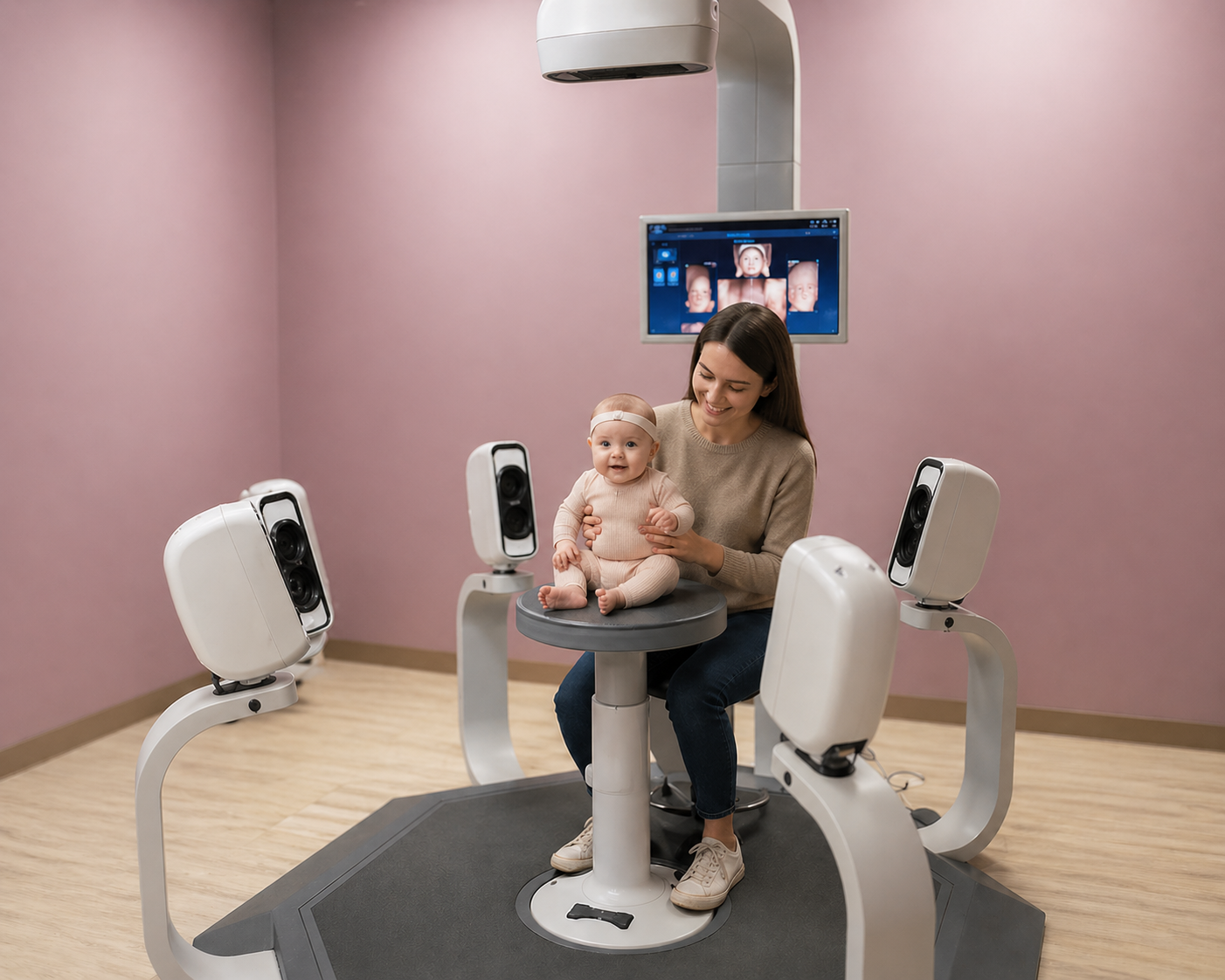
Three-dimensional photography or surface scanning captures the external contour of the head and calculates dimensions digitally. These systems can provide detailed, repeatable surface data but may require specialized equipment, space, training, and an in-person visit. They are not the same as diagnostic imaging of structures inside the skull.
Standardized photography and other validated approaches may also be used. Longitudinal comparison is most interpretable when capture position, landmarks, formulas, and measurement method remain consistent.
Smartphone head shape apps are a newer option. The SoftSpot app, described below, calculates CI and CVAI from guided top-view photos.
Head shape measurement app
SoftSpot® is used by 150+ clinics across the United States
The only FDA-cleared, HIPAA-compliant mobile app for infant cranial measurement, SoftSpot uses a smartphone-based workflow to calculate measurements including CI and CVAI.
FDA-cleared for cranial measurement in infants from 1 month through 2 years, in conjunction with other clinical methods. SoftSpot is not intended for helmet preparation and does not diagnose or prescribe treatment. FDA clearance and HIPAA compliance are separate claims; HIPAA compliance is represented by PediaMetrix.
5. Why timing matters
A retrospective study reviewed 25,332 patients diagnosed with positional plagiocephaly across five Southern California hospitals over 10 years. Compared with infants diagnosed by 6 months, those diagnosed after 6 months were 2.63 times as likely to be referred for helmet evaluation and 1.64 times as likely to require helmet therapy.
This was an observational retrospective study. The findings show an association between later diagnosis and helmet referral or use; they do not prove that earlier detection itself prevents helmet therapy. Clinical differences, access to care, prematurity, hospitalization, and other factors may contribute.
6. Routine head-shape surveillance
AAP guidance on positional skull deformity calls for risk factors to be noted at birth and head shape to be screened during health-supervision visits through the first year. Bright Futures provides the broader framework for age-specific history, physical examination, surveillance, screening, and anticipatory guidance during well-child care.
This does not mean that Bright Futures designates a separate standardized head-shape measurement test at every visit. It means routine pediatric care creates repeated opportunities to examine skull shape and monitor findings. Professionals outside the pediatrician's office may also see infants frequently between visits and can contribute observations, measurements where within scope, and timely communication.
7. Head shape and torticollis
A persistent rotational preference, head tilt, or limited cervical range can concentrate pressure on one area of a growing skull. Positional cranial deformity and congenital muscular torticollis therefore commonly coexist, although either can present without the other.
For PT and OT audiences, relevant observations include active and passive cervical rotation, postural symmetry, tolerance of varied awake positions, motor development, and caregiver handling patterns. The 2024 APTA clinical practice guideline addresses screening, examination, referral, prognosis, and physical therapy management of congenital muscular torticollis. Individualized evaluation—not a generic online protocol—should guide intervention.
Continue supine positioning for every sleep. Position changes and tummy time apply while awake and directly supervised. See the existing torticollis resource for parent-facing context.
8. Tracking head shape over time
Visit 1
Baseline measurement
Visit 2
Repeat measurement
Visit 3
Trend
Longitudinal data can be more informative than memory or photographs alone, which vary with camera angle, scale, hair, and positioning. Record the method, date, infant age, relevant clinical observations, and values so the comparison remains interpretable.
A documented trend can support clearer communication with parents, pediatricians, PTs or OTs, craniofacial specialists, and orthotists when appropriate. No particular numerical change automatically determines treatment.
9. How different pediatric providers may use objective head-shape data
Pediatric PT
Measurement may accompany assessment and management of torticollis, positional preference, posture, and motor development, while providing a consistent head-shape trend for care-team communication.
Pediatric OT
Objective tracking may add context when positioning, movement, participation, handling, or head shape is relevant to the infant's occupational and developmental care.
Lactation / IBCLC
An IBCLC may recognize a head-turn preference or asymmetry during feeding care. Objective information can support neutral communication with the family and appropriate healthcare providers, without implying authority to diagnose a cranial deformity.
Pediatric chiropractic
Objective measurement may document head shape in infants presenting to the practice and support appropriate communication or referral. Measurement is not evidence that chiropractic manipulation treats or corrects plagiocephaly.
Providers should use SoftSpot within their professional scope of practice and applicable state requirements.
10. Communicating with parents
Use neutral, specific language that separates an observation from a diagnosis:
“We noticed some asymmetry in your baby's head shape. We can objectively measure it and track whether it changes over time. These measurements don't provide a diagnosis by themselves, but they can give us useful information to discuss with your pediatrician.”
11. When to refer
Escalate within the appropriate medical pathway for an atypical skull shape, suspected craniosynostosis, progressive or significant asymmetry, significant cervical restriction or torticollis, concerns outside your professional scope, or uncertainty about diagnosis or management. Avoid rigid referral thresholds unless your pathway is supported by an authoritative guideline and applies to the infant and measurement method in question.
Related clinical context is available in the guides to plagiocephaly, brachycephaly, and helmet decision-making.
Craniosynostosis awareness and CranioSpot™
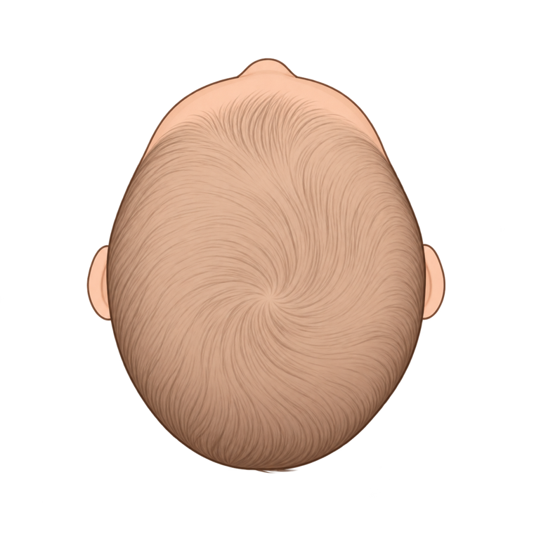
Craniosynostosis results from premature fusion of one or more cranial sutures and requires medical evaluation. CI and CVAI can describe geometry, but they cannot determine whether a suture is fused or exclude craniosynostosis.
CranioSpot™ is a provider-only feature that reviews the same top-down photos collected during a SoftSpot session for head-outline patterns associated with craniosynostosis. Results appear on the provider report, not the parent report.
CranioSpot is not a medical device and is not FDA-cleared for diagnosis or screening. Its recommendation supports clinical judgment; it does not diagnose or rule out craniosynostosis. Pursue atypical findings or clinical concern regardless of a “Within Typical Range” label.
12. FAQ for pediatric providers
How is plagiocephaly objectively measured?
A trained user may compare two standardized diagonal dimensions using cranial vault asymmetry (CVA) or cranial vault asymmetry index (CVAI). The measurement method must be consistent, and results should be interpreted with the history, examination, age, neck mobility, and overall skull shape.
What is CVAI?
Cranial Vault Asymmetry Index expresses the difference between two diagonal cranial measurements relative to the larger diagonal. It describes asymmetry numerically but does not establish a diagnosis or treatment plan.
What is Cranial Index?
Cranial Index, also called Cephalic Index, is head width divided by front-to-back length, multiplied by 100. It describes width-to-length proportion and can help quantify a relatively broad, short head shape.
How often should infant head shape be measured?
There is no universal interval appropriate for every infant or profession. Timing should reflect the clinical question, age, expected growth, measurement method, care plan, and applicable professional guidance. Use a consistent method and document the interval.
Can PTs measure plagiocephaly?
Head-shape observation and measurement may be relevant to pediatric physical therapy, especially when torticollis or positional preference is present. Authority, training, documentation, and billing requirements vary by jurisdiction and setting; PTs should work within their scope and refer when diagnosis or medical evaluation is needed.
Can OTs measure infant head shape?
Objective tracking may be relevant when head shape and positioning affect an infant’s occupational or developmental care. OTs should follow their training, employer policies, professional scope, and applicable state requirements, and should not use a measurement alone as a diagnosis.
Can lactation consultants identify head-shape asymmetry?
An IBCLC may notice asymmetry, head-turn preference, or positioning difficulty during feeding care and communicate those observations. Scope varies, and recognition is not the same as diagnosing a cranial deformity; concerns should be shared with the infant’s pediatric healthcare team.
Can chiropractors measure infant head shape?
A provider may use objective measurement to document head shape if this is within professional scope, training, and applicable state requirements. Measurement does not establish that manipulation will change cranial shape and should support appropriate communication and referral rather than unsupported treatment claims.
Does SoftSpot diagnose plagiocephaly?
No. SoftSpot is FDA-cleared for cranial measurement in infants 1 month through 2 years of age in conjunction with other clinical methods. It does not diagnose plagiocephaly or craniosynostosis.
Can CI or CVAI determine whether a baby needs a helmet?
No. CI and CVAI are descriptive measurements. Helmet decisions require qualified clinical evaluation and consideration of age, diagnosis, severity, growth, prior management, progression, and other findings. SoftSpot is not intended as a tool for preparing a helmet.
When should a baby with head-shape asymmetry be referred?
Refer or escalate when the skull shape is atypical, craniosynostosis is suspected, asymmetry is progressive or clinically significant, cervical restriction or torticollis needs evaluation, the concern falls outside your scope, or diagnosis and management remain uncertain.
Related SoftSpot resources
References
- American Academy of Pediatrics, Committee on Practice and Ambulatory Medicine and Section on Neurological Surgery. Prevention and Management of Positional Skull Deformities in Infants. Pediatrics. 2011;128(6):1236-1241.
- American Academy of Pediatrics. Bright Futures/AAP Recommendations for Preventive Pediatric Health Care: Periodicity Schedule. Updated 2025.
- Congress of Neurological Surgeons. Evidence-Based Guidelines for the Treatment of Pediatric Positional Plagiocephaly. Neurosurgery. 2016.
- Baird LC, Klimo P Jr, Flannery AM, et al. The Role of Physical Therapy. Neurosurgery. 2016;79(5):E630-E631. PMID: 27759674.
- Sargent B, Coulter C, Cannoy J, Kaplan SL. Physical Therapy Management of Congenital Muscular Torticollis: A 2024 Evidence-Based Clinical Practice Guideline. Pediatr Phys Ther. 2024;36(4):370-421. PMID: 39356257.
- Munabi NCO, Nelson MS, Francis SH. Risk Factors for Delayed Diagnosis of Positional Plagiocephaly: A Review of 25,322 Patients. Cleft Palate Craniofac J. 2024;61(10):1679-1686. PMID: 37248557.
- Children's Healthcare of Atlanta. Plagiocephaly clinical documentation and CVAI guidance. 2015.
- U.S. Food and Drug Administration. 510(k) K212045: SoftSpot Cranial Measurement Software. Cleared September 28, 2021.